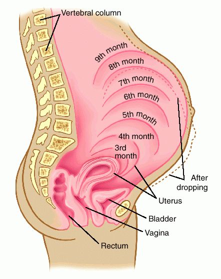
UNSEEN INTERNAL PREGNANCY CHANGES
Your baby’s probable birthday, called your “due date” (or EDC, Expected Date of Confinement) is calculated from the first day of your last normal menstrual period. Classically, a full term pregnancy lasts about 280 days from conception to birth. Full term pregnancy is calculated by 10 lunar (28 day – moon) months or 9 calendar months. Sometimes due dates are calculated based upon implantation bleeding mistaken for a menstrual period. Pregnancy is a time of change in body, heart, mind, and spirit. Relationships change, especially our relationship to yourself. Obvious physical changes include breast changes, growth of the uterus, and a healthy weight gain. Yet during pregnancy your body will go through many unseen invisible changes that help keep you healthy, help maintain pregnancy, and aid in the normal growth and development of your preborn baby (fetus). In the context of relating to your baby before birth, preborn is just a description. Your baby is a baby. Internal Pregnancy Changes During pregnancy, your body goes through many internal unseen changes including: Formation, implantation, & growth of the placenta; Maternal blood volume expands 40-60%; Maternal liver increased function demand; Growh of the uterus; Breast changes initiate lactation. The Placenta The human placenta is the tree of life to a preborn baby. Formation of the Placenta The placenta is a fetal organ belonging to a preborn baby. The placenta begins to form about 11 days after conception. When conception occurs in a fallopian tube, the fertilized ovum (egg) floats through the fallopian tube to the inside of the uterus where it burrows into the lining of the uterus, in a process called implantation. The process may take three or four days. Implantation After six or seven days the fertilized ovum, now called a blastocyst, is ready to embed inside the uterus. Tiny root-like projections with the ability to break down tissue (called syncytiotrophoblast) invade the endometrial lining of the uterus. A chemical reaction within the uterus (decidual reaction) limits the invading capacity of the embryo. This process of implantation is complete when the endometrial cells heal over the opening enclosing the embryo. During the third week of pregnancy chorionic villi (chorionic frondosum) completely covering the embryo penetrates blood vessels they touch and are bathed in pools of maternal blood. The open areas around the blood vessels are called sinuses and the areas of maternal blood surrounding the villi are known as blood spaces. By the day 13 after conception the entire blastocyst is covered with trophoblastic cells. The blastocyst forms rudimentary chorionic villi (placental tissues not yet containing blood vessels). Rudimentary chorionic villi absorb nutrients from broken down tissue in the implantation cavity. The placenta grows as pregnancy progresses. Poor implantation of the placenta may be associated with malnutrition in early pregnancy. Two Sides of the Placenta Maternal Side of the Placenta The maternal side of the placenta is made of chorionic villi called cotyledons (or lobes) which are separated by furrows. Cotyledons fit together like pieces of a puzzle, and their surface is covered with trophoblastic cells. The maternal side of the placenta is bluish-red color. Fetal Side of the Placenta The fetal side of the placenta is white, smooth, and shiny. Branches of the umbilical vein and arteries and insertion of the umbilical cord can be seen. The fetal side of the placenta is covered with two membranes called the chorion and the amnion. The chorion and amnion continue beyond the outer edge of the placenta forming the amniotic sac containing the amniotic fluid and preborn baby. All of the blood flowing through the placenta is preborn baby’s blood circulating between the placenta and preborn baby through the umbilical cord. The placenta grows during pregnancy to keep up with the nutritional demands of the growing and developing preborn baby. What Does the Placenta Do? The placenta is a fetal organ belonging to a preborn baby. The placenta is born after a baby’s birth during the third stage of childbirth. Also called the afterbirth, the placenta functions as the respiratory, digestive, excretory, and eliminatory systems of a preborn baby. The placenta transfers oxygen and nutrients from mother to preborn baby and waste products from preborn baby to mother. Preborn babies make breathing movements and are not breathing using their lungs. Oxygen is obtained from hemoglobin in a mother’s blood by diffusion from the maternal circulation through the placenta into the fetal circulatory system. A preborn baby gives off carbon dioxide into maternal blood through the placenta in a process much like obtaining oxygen. The excretory process results from a catabolic (breaking-down) metabolic process. Preborn baby’s excretory process is minimal because the metabolism is anabolic (building-up). Malnutrition in early pregnancy is associated with poor implantation of the placenta. Poor implantation of the placenta includes conditions such as placenta previa, placenta accreta, and low lying placenta. Each of these complications of the placenta are associated with complications of pregnancy. Malnutrition during pregnancy is very different than malnutrition when you’re not pregnant. The nutritional demands of pregnancy are that you eat a daily diet of fresh, wholesome, real foods. Digestive The placenta transfers oxygen, nutrients, and all other substances a mother ingests to preborn baby (fetus). According to Dr. Thomas H. Brewer, a preborn baby is not a nutritional parasite. In order for a preborn baby to receive nutrients through the placenta, a pregnant mother has to eat first! Your baby is what you eat! You are building your preborn baby’s body! It has to last a lifetime, build a great one! Excretory The excretory process results from a catabolic (breaking-down) metabolic process. Preborn baby’s excretory process is minimal because the metabolism is anabolic (building-up). A preborn baby gives off carbon dioxide into the maternal blood through the placenta in a process much like obtaining oxygen. Respiratory The respiratory function of the placenta provides oxygen to a preborn baby. Oxygen is obtained from hemoglobin in maternal blood by diffusion from the maternal circulation through the placenta into the fetal circulatory system. Preborn babies make breathing movements and are not actually breathing using their lungs while in the womb. There are no pulmonary exchanges of gases while a preborn baby is in the womb. The placenta grows as pregnancy progresses until about the 35th week of pregnancy. Maternal Blood Volume Maternal blood volume increases as pregnancy advances to full term. Maternal blood volume eventually increases 40 to 60 percent above non-pregnant circulation. Increase in the amount of blood in the circulation of a pregnant mother is to service the placenta in the exchange of nutrients, gases, and other substances between a mother and her preborn baby. A benefit of increased maternal blood volume is that fluid is stored as a protection against shock in case of excess blood loss during childbirth. Growth of the Uterus During pregnancy the uterus grows thirty times larger than its non-pregnant size. A pregnant uterus must grow to accommodate the growing placenta, amniotic sac and fluid, and the preborn baby. Rapid growth of the uterus during pregnancy is made possible by connective tissue called collagen. Strength of the uterine contractions during labor is a nutrition-related function. Maternal Liver The liver has more than 500 metabolic functions in the human body. During pregnancy, three functions are very important. According to “Preventing Nutritional Complications of Pregnancy” by Dr.Thomas H. Brewer, these three liver functions are: “1. Albumin Synthesis One of the most complicated processes the liver governs involves the selective combining of specific amino acids into protein molecules which serve to maintain an appropriate amount of fluid in the bloodstream. When the liver is damaged, albumin synthesis is one of the first functions affected. If albumin levels in the bloodstream drop, water, which should be in the circulation leaks out into the tissues. This causes abnormal swelling and puffiness (pathologic edema) and leaves the blood volume contracted. 2. Hormone Metabolism The liver clears from the body a staggering load of female hormones manufactured continually by the placenta. The level of increased hormonal activity is equivalent to that of 100 birth control pills a day. Proper metabolism of these hormones requires that the liver attach the fat-soluble hormones to other molecules which make them water soluble, then excrete them in the bile or return them to the circulation, whereby they are excreted by the kidneys in the urine. If the liver falls behind in this function, hormones can back up in the tissues and reach toxic levels. 3. Detoxification Following a pattern similar clearing hormones, the liver metabolizes toxins entering the bloodstream from the lower bowel. Digestive processes slow during pregnancy, giving toxins a friendlier environment to develop, thus increasing the stress on the liver.” (*) Breast Changes Initiate Lactation As soon as you conceive a baby, your body begins to prepare for lactation, even if you don’t plan to breastfeed. By the time you are ready to give birth, your body is ready to nourish your baby by producing milk. During pregnancy, milk glands and milk ducts mature and begin to produce colostrum, the pre-milk available to baby for around 3 to 5 days before true milk comes in. ** **Adapted and/or reprinted with written permission of the author, Thomas H. Brewer, M.D. (c) 1978, Preventing Nutritional Complications of Pregnancy, page 5, Society for the Protection of the Unborn through Nutrition (SPUN) |
Thank you in advance for your donationPayPal is safe and secure
"Doctor Thomas H. Brewer , author of "Metabolic Toxemia of Late Pregnancy" wanted pregnant women and those working with them to think of pregnancy from the inside out." The Human Placenta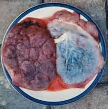
Two human placenta showing the maternal side (left) and the fetal side with umbilical cord and amniotic membranes.
The maternal side of the placenta is made of chorionic villi called cotyledons (or lobes) which are separated by furrows. The fetal side of the placenta is white, smooth, and shiny. Healthy Mothers make Healthy Babies! Save a Preborn Baby! Feed a Pregnant Mother! |
| Auntie Natal Educational Services |
|
